AMA vs Medicare rules and the use of the PT modifier

AMA vs Medicare rules and the use of the PT modifier

By:  Chris Woolstenhulme, QCC, CMCS, CPC, CMRS
Published:  May 22nd, 2018

Be sure to review the specific payer policy you are submitting claims to. Medicare’s policy requires the use of a different code when a screening colonoscopy becomes a diagnostic procedure requiring you to bill with CPT code 00811 when treating a Medicare Beneficiary. The use of the PT modifier is also a Medicare rule, see information below from the WPS website.

With that being said, what I am saying is if the payer you are billing has a policy and the provider is contracted with that payer, the provider is obligated to follow the payer’s policy. AMA does indeed have guidelines, but a payer may have their own rules, and if so you need to follow the payer’s rules. A lot of payers follow CMS policies and rules such as BC, OPTUM, UHC…. In regards to reaching our to your payer, the claims department will likely not be of help, However, most providers are assigned a Provider Representative that is there for the provider and staff, you should know your large provider Reps by their first name.

The key is to UNDERSTAND the specific payer policies and the rules you need to follow for each payer. The AMA may have guidelines; however, your contract with specific payers may trump those guidelines. Find-A-Code has access to Commercial Payer policies you may be interested in; it is one of our most popular tools. You will improve your bottom line and ensure compliance if you have someone managing your contracts that can identify and understand the specific rules, as well as have payer policies easily available.

Don’t forget the date of Service, was the service done before particular codes went into effect? Rules may change with payers and you are expected to keep up with them and comply with the changes on the effective date of the change. In the case of an audit, payers will look at the contracts the provider has signed with them and expect compliance with their rules.

PT Modifier

Definition: A colorectal cancer screening test which led to a diagnostic procedure

Appropriate Usage:
When a service began as a colorectal cancer screening test and then was moved to diagnostic test due to findings during the screening. Practitioners should append the modifier to the diagnostic procedure code that is reported instead of the screening colonoscopy or screening sigmoidoscopy HCPCS code
Append to procedure codes in the range: 10000 to 69999

Inappropriate Usage:
Do not use the Modifier PT when the service began as a diagnostic procedure
On any other procedure outside the range listed above

References:

CMS Medicare Learning Network Matters Article MM7012
Note: The Medicare policy is that the deductible is waived for all surgical procedures furnished on the same date and in the same encounter as a colonoscopy, flexible sigmoidoscopy, or barium enema that were initiated as colorectal cancer screening services.

WPS – Government Health Administrators- Modifier PT Fact Sheet

As per CMS MLN- MM10181:

00811 – Anesthesia Diagnostic Colo (4 base units)

00812 – Anesthesia Screening Colo (3 base units)

Anesthesia services furnished in conjunction with and in support of a screening colonoscopy are reported with CPT code 00812 (Anesthesia for lower intestinal endoscopic procedures, endoscope introduced distal to duodenum; screening colonoscopy). CPT Code 00812 will be added as part of January 1, 2018, HCPCS update.

Effective for claims with dates of service on or after January 1, 2018, Medicare will pay claim lines with new CPT code 00812 and waive the deductible and coinsurance. When a screening colonoscopy becomes a diagnostic colonoscopy, anesthesia services are reported with CPT code 00811 (Anesthesia for lower intestinal endoscopic procedures, endoscopy introduced distal to duodenum; not otherwise specified) and with the PT modifier. CPT code 00811will be added as part of January 1, 2018, HCPCS update.

Effective for claims with dates of service on or after January 1, 2018, Medicare will pay claim lines with new CPT code 00811 and waive only the deductible when submitted with the PT modifier.

References:
What Do We Offer Our Attorney Clients?

We offer years of nursing experience.

Nurses are more detail oriented; we are trained to assess the details.

We pick up on the details an MD may miss because they are more about the “big picture.”

We are more cost effective than an MD who charges much more to review/analyze medical records.

We can tell you the exact expert that you need to support your case in trial.

Call us at 618.376.3089 today for a free consultation. Have a great day!


Pneumonic Plague

The case of a man in Colorado who contracted human pneumonic plague in June 2014 was published today by the Centers for Disease Control and Protection (CDC). This case was unusual in that plague only affects around eight people annually in the United State, and that the source of the infection was his dog.

Pneumonic plague is a rare but life-threatening condition caused by the bacteriaYersinia pestis. Y pestis is usually carried by rodents and can be transmitted to humans through a bite from an infected rodent or rodent flea or inhalation of infectious droplets. However, it usually develops as a complication of untreated bubonic plague. Pneumonic plague is generally fatal if not treated with antibiotics and is the only form of plague with the potential for human-to-human transmission.

The Colorado patient, a previously healthy middle-aged man, was admitted to a local hospital after a fever and cough became rapidly worse. He was diagnosed with pneumonia and P luteola was identified as the causative agent by an automated identification system. Despite treatment, his condition continued to deteriorate so his samples were reanalyzed manually and Y pestis was identified. The man recovered after treatment with broad-spectrum antibiotics.

It transpired that a few days before the man had become ill, his dog had been put down due to respiratory difficulties. Analysis of samples from the dog confirmed that it too was infected with Y pestis. The veterinary clinic confirmed that the man had been in prolonged close contact with his dog during the last hours of its life.

Two veterinary nurses who had cared for the dog and a friend of the Colorado patient also developed fever and respiratory symptoms and tested positive for Y pestis. All three subsequently made a full recovery. It is not known whether the friend contracted the plague from the dog or the Colorado patient.

This is the largest plague outbreak in the United States since 1924, and could also include the first case of human-human plague transmission.

As a result of this outbreak awareness needs to be raised regarding the potential human risk of sick domestic animals. Furthermore, it has drawn into question the use of automated culture identification systems since the initial incorrect diagnosis of the Colorado patient unnecessarily exposed additional medical staff to the plague.

Source:

Runfola JK, et al. Outbreak of Human Pneumonic Plague with Dog-to-Human and Possible Human-to-Human Transmission — Colorado, June–July 2014. Morbidity and Mortality Weekly Report (MMWR) May 1 2015;64(16):429–434.

Drug News: Zyprexa Study

Zyprexa is used to treat schizophrenia to improve symptoms which include hearing voices, seeing things that are not there, and being suspicious and withdrawn. The label carries a “boxed warning”, the FDA’s most serious type of warning, for post-injection delirium sedation (PDSS). This is an update to the MedWatch safety alert of June 18, 2013. http://www.fda.gov/Safety/MedWatch/SafetyInformation/SafetyAlertsforHumanMedicalProducts/ucm357601.htm

The FDA concluded a review of a study undertaken to determine the increased levels of injectable Zyprexa in two patients who died. The study results were inconclusive and the FDA was unable to exclude the possibility that the deaths were caused by rapid, but delayed, entry of the drug into the blood stream following intramuscular injection. Much of the drug increase could have occurred after death. This finding could explain the extremity high blood levels found int the patients who died 3-4 days after receiving the appropriate doses of the drug. After reviewing all the information, the FDA does not recommend any changes to the current prescribing or use of Zyprexa Relprevv injection at this time.

Patients should review the medication guide/insert each time they received an injection because there may be new information.

If you come across any Zyprexa injuries in your case load:

  • Be sure to analyze all the symptoms reported in the medical records.
  • Use the above link to the safety article as a reference.
  • Research articles on Zyprexa problems, issues, and adverse reactions.
  • Consult with an RN if you have any questions or need any assistance.

If we can be of assistance on any of your medically related cases, please call us at 618-376-3089 or 618-401-0746.

Until next time…

Personal Injury: Traumatic Brain Injury (TBI)

What is traumatic brain injury? TBI is a common injury which is easily missed when the trauma team is focused on saving an individual’s life. The effects of TBI are significant.

There are two categories of TBI: Mild and severe. Mild TBI is diagnosed as injury with loss of consciousness and/or confusion/disorientation which is shorter than 30 minutes. In mild TBI, a CT scan or MRI are often seen as normal while the individual has cognitive problems, headache, difficulty concentrating and thinking, memory problems, attention deficits, mood swings and frustration. Even mild TBI can be devastating to the injured person and their family.

Severe TBI is associated with loss of consciousness longer than 30 minutes and memory loss after the injury lasts longer than 24 hours. Deficits range from impairment of higher level cognitive functions to comatose states. Survivors may have limited function of extremities, abnormal speech, loss of ability to think or emotional problems.  Survivors with severe injuries can be left in a long-term unresponsive state. Long-term rehabilitation may be required.

There are many injuries that can cause TBI. These are the mechanisms that are the highest causes: Open head injury, closed head injury, deceleration injuries, chemical/toxic, hypoxia, tumors, infections and stroke. Open head injury would be something like a gunshot wound that causes penetration of the skull. Closed head injury would be something like a slip and fall and vehicular accidents for example. Deceleration injuries is what is caused when the skull is moving fast and then suddenly stops, banging the brain against the skull. Shaken baby syndrome is another example.

Diffuse axonal shearing: when the brain is slammed back and forth inside the skull it is alternately compressed and stretched because of the gelatinous consistency.  The long, fragile axons of the  neurons (single nerve cells in the brain and spinal cord) are also compressed and stretched.  If the impact is strong enough, axons can be stretched until they are torn.  This is called axonal shearing.  When this happens, the neuron dies.  After a severe brain injury, there is massive axonal shearing and neuron death.

Chemical/toxic injury occurs when harmful chemicals damage the neurons. These chemical can include insecticides, solvents, carbon  monoxide poisoning and lead poisoning. Hypoxia means lack of oxygen to the brain. This causes irreversible brain injury. Infections such as meningitis and encephalitis can cause TBI. Stroke is another mechanism of TBI, cell death can occur from lack of oxygen to the brain cells caused by the stroke.

If you practice personal injury cases, you may well have worked on these types of cases.

Plaintiff Attorneys should look for the following information:

  • Was there failure to diagnose, failure to timely treat or transfer to a higher level of care?
  • Did staff document and consider the mechanisms of injury?
  • Was a head injury suspected?
  • Were staff trained in the specific knowledge of trauma and follow the standard of care?
  • Where did the injury occur?
  • When did the injury occur? Timing is important; time from injury to first responder’s care.
  • Was the treating facility a trauma center or a rural facility?

Defense Attorneys should look for the following:

  • Were there any pre-existing conditions that could contribute to the head injury complications?
  • Was there adequate staff in place?
  • What else was going on in the ER at that time? Confusion due to a full house?
  • If there were severe pre-existing conditions, was the patient compliant with his/her care?
  • Does the patient share in the negligence in any way?
  • Was there alcohol or illicit drug use involved?

How to Interpret an EKG Strip – Continuing with the next step – PR Interval

EKG-Printerval

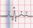
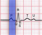
The PR interval indicates AV conduction time. In this step you should measure the interval from where the P wave begins until the beginning of the QRS complex. Calipers, marked paper or counting small boxers methods can be used to determine PR intervals. Normally this interval is 0.12 to 0.20 seconds (3 to 5 small boxes) in adults, longer in the elderly. This interval shortens with increased heart rate.

Also evaluate if PR intervals are constant or varying across the EKG strip. If they vary, determine if the variations are a steady lengthening until the point where an expected QRS does not appear.

PR interval questions to address:

  1.  Does the PR interval fall within the norm of 0.12-0.20 seconds?
  2.  Is the PR interval constant across the EKG tracing?

QRS Complex

EKG-QRSinterval


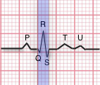
The QRS complex indicates ventricular depolarization. Depolarization triggers contraction of the ventricles. Because of the larger tissue mass, the QRS complex is larger than the P wave. While the prototypical QRS complex consists of three wave components, one or two of these components may be missing. In this step, measure the QRS interval from the end of the PR interval to the end of the S wave. Use calipers, marking paper or by counting small boxes. normally this interval is 0.06 to 0.12 (1.5 to 3 boxes).

QRS questions:

  1. Does the QRS complex fall within the range of 0.06-0.12 seconds?
  2. Are the QRS complexes similar in appearance across the EKG tracing?

T Wave

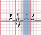
EKG-TThe T wave indicates the repolarization of the ventricles. It is a slightly asymmetrical waveform that follows (after a pause) the QRS complex. Take note of the T waves that have a downward (negative) deflection or of T waves with tall, pointed peaks. The U wave is a small upright, rounded bump. When observed, it follows the T wave.

QT Interval

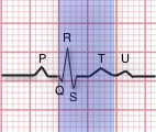
EKG-QtintervalThe QT interval represents the time of ventricular activity including both depolarization and repolar- ization. It is measured from the beginning of the QRS complex to the end of the T wave. Normally, the QT interval is 0.36 to 0.44 seconds (9-11 boxes). The QT interval will vary with patient gender, age, and heart rate. Another guideline is that a normal QT interval is less than half of the R-R interval for heart rates below 100 beats per minute.

ST Segment

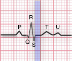
EKG-StsegmentThe ST segment represents the early part of ventricular repolarization. The ST segment is the line that from the end of the QRS complex to the beginning of the T wave. Normally the ST segment is flat relative to the baseline.


I hope this has helped you understand what the EKG strip is showing.

Take care and see you next time!


How to Interpret an EKG Strip – Continued

EKG interpretation should be performed using a standard procedure, such as this eight step procedure:

  • Rhythm
  • Rate
  • P Wave
  • PR Interval
  • QRS Interval
  • T Wave
  • QT Interval
  • ST Segment

EKG-regular

EKG-irregular


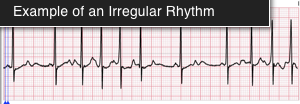
For ventricular rhythms, examine the R to R intervals on the EKG strip. Calipers or paper marks can be used to fix the distance for R-R interval and then this distance can be compared to other R-R pairs.  Are they regular, meaning that each heart beat’s R-R interval is equal? Small variations of up to 10% are considered equal. Is the rhythm regularly irregular? For example, is there a  pattern, such as increasing R-R durations? or perhaps groups of similar intervals as illustrated, or are the R-R intervals completely irregular.

For atrial rhythm, observe the P-P intervals. Are they regular (minor variations can be caused by the breath cycle)? If P-P intervals are irregular, is there a pattern?

EKG-HR1


EKG-HR2


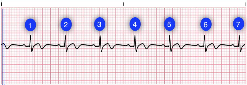
There are several methods for determining heart rate. The first method is simple. Count the number of QRS complexes over a 6 second interval and multiple by 10 to determine heart rate. In the first image, we can count 7 QRS complexes, so the heart rate is 70.

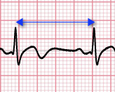
The second method uses small boxes. Count the number of small boxes for a typical R-R interval. Divide this number into 1500 to determine the heart rate. In the second image, the number of small boxes for the R-R interval is 22.5. The hear rate is 1500/21.5 = 69.8 beats per minute.

EKG-P


The P Wave represents atrial depolarization. In a normal EKG, the P-wave precedes the QRS complex. It looks like a small bump upwards from the baseline. The amplitude is normally 0.05 to 0.25 mV (0.5 to 2.5 boxes). Normal duration is 0.06-0.11 seconds (1.5 to 2.75 small boxes). The shape of a P-wave is usually smooth and rounded.

Let’s leave it here for now and next time we will pick up with the PR Interval.

I want to give you time to digest this section before moving on.

How to Read an EKG Strip – The EKG Paper: What Does it Mean?

How to Read an EKG Strip – The EKG Paper: What Does it Mean?


If you work in the medical or legal field, you’ve probably already seen an EKG strip and you know what it looks like.

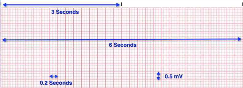
The EKG tracings are recorded on grid paper. The horizontal axis shows time and the vertical axis records EKG amplitude or voltage of the impulse.

EKG-grid1

The black marks at the top of the paper indicate 3 second intervals as seen above. Each 5 squares equals one second, so there are 15 squares in the 3 second interval on the paper. That means that each large square equals 0.2 seconds.

Regarding the vertical lines indicating the amplitude, each 2 large squares equals 1 millivolt; each small block equals 0.1 mV. Within the large blocks are 5 small blocks, representing 0.04 second each.

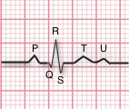
Normal EKG tracings consist of waveform components that indicate electrical events during one heartbeat. These wave-forms are labeled P, Q, R, S, T and U.

EKG-oneBeat

The P-wave is the first deflection and is normally positive or upward. It indicates atrial depolarization.

The QRS complex follows the P-wave. A normal QRS complex is indicated above; a short downward deflection (Q) followed by a sharp upward deflection (R) and then a downward deflection (S) S-wave. The QRS complex represents ventricular de-polarization and contraction.

The T-wave is normally a modest upward wave form, representing ventricular re-polarization. The U-wave indicates the recovery of the Purkinje conduction fibers. This wave component may not always be observable.

This will be continued in the next blog. Stay tuned!

Part 2: EKG Interpretation.

What is Schizophrenia?

Schizophenia is a mental disorder that generally appears in late adolescence or early adulthood, however it can emerge at any time in life. It is one of many brain diseases that may include delusions, loss of personality (flat affect), confusion, agitation, social withdrawal, psychosis, and bizarre behavior.

Schizophrenia is a chronic, severe, and disabling brain disorder that has affected people throughout history. Those affected may hear voices. They may believe that others are trying to read their minds, control their thoughts, or are plotting to hurt them in some way. This can terrify sufferers and make them withdrawn or very agitated. They have trouble holding a job or caring for themselves, so they rely on others for assistance.

My deceased brother-in-law was diagnosed with Schizophrenia. As long as I knew him he was affected. He would hear voices and think that others were talking about him when they were not. He suffered from delusions and paranoia. It is a heart-wrenching disease when one has to watch a loved one endure it. He was diagnosed early in life with childhood schizophrenia.

There is often a phase leading up to the disease, called premorbid or prodromal. This phase is more pronounced in children than adults. In children, the premorbid impairments include: language impairments, motor (movement) effects, and social deficits. Experts think that causes include genes, environment, and different brain structure and chemistry. It affects about 1% of the population.

Schizophrenia is difficult to diagnose. There is no one test to determine it. It takes a psychiatrist or psychologist to analyze the person to determine whether there is psychosis present.

Symptoms include hallucinations, delusions, thought disorders, movement disorders, flat affect, speaking very little, lack of pleasure in everyday life, cognitive symptoms.

Treatments include antipsychotic medications such as Thorazine, Haldol, Trilafon, Prolixin, Risperdal, Zyprexa, Seroquel, Geodon, Abilify, and Invega. When working on a case with a diagnosis of schizophrenia, be sure to look for these medications in the medical records to confirm whether your client is being treated for the disease.

For further information, check out the National Institute of Health, The BBR Foundation, The Breakaway Study, and the Mayo Clinic.

Update

Hello everyone,

A quick update for you; I just returned from Asheville, NC from a two-day class, the final lesson in the life care planning program I am taking through Fig Services. I have one more task to complete and by spring I should be ready to offer life care planning services to my attorney clients on their catastrophic cases, such as spinal cord injuries and traumatic brain injuries.

I am looking forward to this new challenge and my goal is to be able to assist attorneys with these type of cases. It will be a huge benefit to them, so I’m really looking forward to helping.

I will resume the educational pieces next week. Thank you for following! Have a great week!

Different Types of Spinal Disk Problems

Different Types of Spinal Disk Problems

By Pam Steinacher, RN, LNC, MSCC, CHRM

This blog will show disease of the spinal vertebrae and disks. These photos may come in handy when you are working on a case in which spinal surgery has been performed. The images below show different types of disk disease and degeneration. Whether you are plaintiff or defense, this blog can make things a little clearer in your mind.

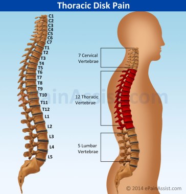
There are seven cervical vertebrae, twelve thoracic vertebrae, and five lumbar vertebrae in the human spine. There is also a sacrum, which normally has five bones in it. We are only concerned today with the spinal column and its 24 vertebrae.

human spine

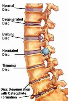
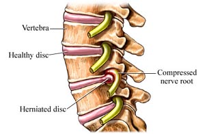
The below image shows the normal disk versus a degenerated, bulging, herniating, thinning, and degenerated disk. The disk degeneration with osteophyte (bone spur).

disc-problems

Below is a pinched (compressed) nerve root. Pinched nerves can be a source of severe pain.

compressed_nerve

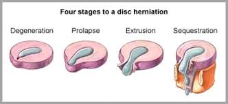
This image shows the four stages to a disk herniation. It starts out as degeneration of the nucleus pulposus, and continues to the prolapsed stage. Then the nucleus extrudes from the disk and then finally sequestration of the disk.

4 stges to disk herniation

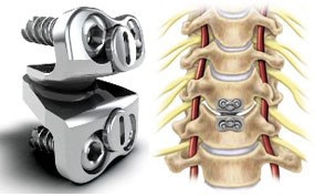
This is one type of hardware used in spinal fusion surgery.

hardware for fusion

I hope you find this blog useful in your practice. Until next time…

Pam

P.S. I am working on the layout to this blog. For some reason I cannot get it to post correctly. Thank you for your patience.

Finding the Perfect Expert Witness for Your Case

Finding the Perfect Expert for Your Case

By Pam Steinacher, RN, LNC, MSCC, CHRM


One of the services I provide as a legal nurse consultant is locating experts for attorneys to support the issues of their case. This can be a challenging task as there are several factors involved in locating the best expert to support and strengthen your case.

I must first discuss with the attorney the facts of the case. It is most helpful if I can review the medical records first in order to locate the exact expert needed, so that I can compare “apples to apples” so to speak.  I can locate experts for liability, standard of care, causation, damages and the fact witness expert and any other type you may need.

Once the type of expert is established, I then put out “feelers” among my database of experts to see who is available and appropriate. I then determine which one of those available is the perfect one for your case. For example, if your case involves a medication administration error I would first have to establish what type of staff member and what type of facility was involved. Different facilities have different protocols directing mediation administration. Hospitals have different rules than long term care facilities and each have their own policies.

Location is also an issue. I strive to find an expert as close to the location of the case as possible in order to keep the attorney’s costs down regarding the expert’s travel expenses. I am always trying to look out for my attorney client’s wallet.

Some attorneys like to vet the expert themselves or I can do it for you. I am always trying to meet my attorney client’s needs. By providing the vetting service, it saves my attorney client time in his or her busy schedule.

When you need an expert for your medically related case, call me at 618.376.3089 and I will be happy to discuss your case with you and determine the best expert witness to strengthen your case.


Pam Steinacher, RN, LNC, MSCC, CHRM

Steinacher Medical Legal Consulting

www.steinachermedlegal.com/

pam@steinachermedlegal.com

618.376.3089

Deciphering Medical Records

Steinacher Medical Legal Consulting’s Blog

By Pam Steinacher, RN, LNC, MSCC, CHRM

Do you sometimes have trouble reading medical records? Can you decipher this?

Hand written progress note


This note says Antivert 12.5 mg. If you had trouble with this, you are not alone. Many attorneys and paralegals have difficulty deciphering medical records, especially handwritten records. They are the worst! We all know that doctors have the worst handwriting of all the professions.

Here’s another one, what does this say?

Written progress notes


Did you get “Called requesting soft restraints, I told her I would prefer chest restraints at this time” out of it? That’s what it says. Would you have needed help to figure it out.

Nurses work with this type of thing all day long and have become very proficient at deciphering doctors’ handwriting. We have to be able to read the progress notes and orders in order to carry them out in a timely manner.

When you have trouble with medical records, you need a nurse consultant on your side.

Steinacher Consulting Blog

Why Should I Hire Legal Nurse Consultant?

by Pam Steinacher, RN, LNC, MSCC, CHRM, Legal Nurse Consultant

Legal Nurse Consultants (LNCs) are an added resource for the paralegal. LNCs work with the paralegal in analyzing a case and reviewing medical records. The paralegal knows the legal process and the LNC, or RN, knows the medical issues and can educate on medical terms and medical conditions. A paralegal can become very proficient at reading medical records over time, but can s/he interpret those records? Do the attorney and paralegal know what the medical terms mean? Do the attorney and paralegal know what the different lab values mean? Or why they are important? Does the attorney know what records should be in a chart and tell if something is missing? A good LNC can help with all of these questions.

There are many different ways in which an LNC can assist on medical, injury or death cases. To name a few:

  • Educating attorneys regarding medical facts and issue relating to the case or claim;
  • Integrating nursing knowledge with the challenge of criminal and civil litigation;
  • Assisting the acquisition of records and other medically related materials;
  • Preparing chronologies of medical events and correlating them to the allegations;
  • Identifying deviations in the standards of care and damage issues;
  • Helping to determine the merit of a case;
  • Screening for record tampering;
  • Conducting research and summarizing medical literature;
  • Attending, witnessing, and reporting on independent/defense medical exams;
  • Assisting with depositions, trials, and alternative dispute resolutions, including developing and preparing persuasive graphic exhibits;
  • Preparing witness and exhibit lists;
  • Conducting client interviews.

Because of the LNC’s expertise in medically related issues, the s/he can bring the following benefits to your litigation team:

  • Experience; our background of clinical experience includes the interpretation of medical records, documents and medical-legal issues;
  • Cost effectiveness; we are cost effective because we can evaluate charts more efficiently since we know the standards of care, language and expectations of care (we can identify the strengths and weaknesses of of the case because we understand the medical issues at hand);
  • Resourcefulness; we possess a network of medical and professional resources and contacts;
  • Knowledge; we have a thorough understanding of medical issues and how they apply to the litigation process. This information is custom designed to the attorney, client, and jury.

Attorneys nationwide are slowly finding how an LNC can be crucial to a positive case outcome.

Testimonial:
Steinacher Medical Legal Consulting finds our hard to find expert witnesses for us without fail. Experts are located in a timely manner and are just what we need to support our case.

Veronica Sheffa, Legal Assistant
Patrick Salvi, Partner
Salvi, Schostok & Pritchard

Ms. Steinacher has been working with attorneys nationwide since January 2004 reviewing cases and assisting to improve successful outcomes. She is the president/owner of Steinacher Medical Legal Consulting. She can be reached at 618.376.3089 for a complimentary case consultation.